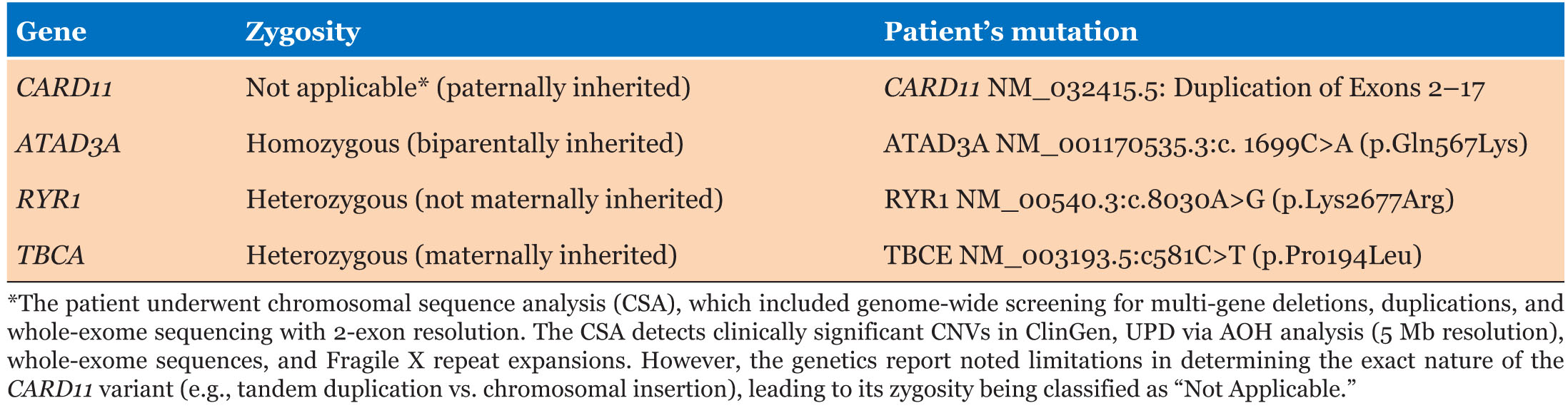
|
Case Report
Gastric teratoma: An uncommon cause of gastrointestinal bleeding in children
1 University of Toledo College of Medicine and Life Sciences, Toledo, OH, USA
2 Department of Pediatrics, Nationwide Children’s Hospital, Columbus, OH, USA
3 Department of Pediatric Gastroenterology, ProMedica Russell J. Ebeid Children’s Hospital, Toledo, OH, USA
Address correspondence to:
Jenny Chen
3000 Arlington Avenue, Toledo, OH 43614,
USA
Message to Corresponding Author
Article ID: 100014Z19JC2022
Access full text article on other devices

Access PDF of article on other devices


How to cite this article
Chen J, Springer J, Banerjee S, Kumar R. Gastric teratoma: An uncommon cause of gastrointestinal bleeding in children. J Case Rep Images Pediatr 2022;4:100014Z19JC2022.ABSTRACT
Introduction: Gastric teratomas are a rare cause of abdominal mass in the pediatric population.
Case Report: We report a 26-day-old male who presented with hematemesis. Initial abdominal radiography showed a round soft tissue density within the stomach despite unremarkable abdominal ultrasonography. Upper endoscopy revealed a polypoid mass within the fundus of the stomach without active bleeding. Computed tomography showed a hypodense area in the gastric fundus without exo-gastric involvement. Surgical excision by laparotomy was successfully completed with the excision of a 3.2 × 2.0 × 1.4 cm polypoid mass with a focal hemorrhagic surface, arising from the posterior wall of the stomach. Histopathology of frozen section confirmed the diagnosis of a benign mature teratoma of the stomach. Post-operative course was uneventful. Six-month follow-up upper endoscopy biopsies were grossly normal with no evidence of tumor reoccurrence.
Conclusion: Gastric teratomas have a favorable prognosis but require a high index of suspicion for diagnosis due to its variable and non-specific presentation.
Keywords: Endo-gastric tumor, Gastric teratoma, Mature, Pediatric
Introduction
Gastric teratomas are rare tumors in the pediatric age group. As supported with our case, the vast majority of gastric teratomas present in children under one year of age and have a predominance for males. Although 60% are exo-gastric in nature, our report discusses an endo-gastric presentation which accounts for only 30% of gastric teratomas [1]. Due to non-specific symptoms and a broad differential diagnosis for abdominal masses, pre-operative diagnosis can be difficult to ascertain. However, with complete surgical excision, gastric teratomas have a good prognosis. We report a case of an infant presenting with an endo-gastric benign mature teratoma.
Case Report
A 26-day-old male presented to the emergency room with complaint of hematemesis. He was anemic but otherwise had an unremarkable physical examination. Work up demonstrated hemoglobin 6.4 g/dL and normal international normalized ratio. He was kept nothing by mouth and admitted to inpatient pediatric care. Abdominal ultrasonography was unremarkable, but abdominal radiography showed a round soft tissue density within the stomach which prompted upper endoscopy evaluation. Initial endoscopy noted a polypoid mass within the fundus of the stomach without active bleeding (Figure 1). No biopsy of the mass was taken at the time due to risk of bleeding. Differential diagnosis after endoscopy included malignancy, hamartomatous or hyperplastic polyp, teratoma, vascular lesion, or other rare tumors. A computed tomography was obtained to evaluate the extent of mass invasion which revealed a hypodense area in the gastric fundus without exo-gastric involvement. Pediatric surgery was then consulted for excision of the mass.
Surgical excision by laparotomy was completed with successful excision of a 3.2 × 2.0 × 1.4 cm polypoid mass, embedded within the gastric wall and arising from the posterior wall of the stomach with a focal hemorrhagic surface. The final pathological diagnosis made by frozen section was a pedunculated polypoid benign mature teratoma of the stomach with an ulcerated inflamed surface. The patient’s post-operative course was uncomplicated. His hemoglobin remained stable, and he was tolerating formula. He was discharged home with close follow-up. An endoscopy scheduled six months later for follow-up was grossly normal (Figure 2). There was no evidence of tumor reoccurrence, and the patient was gaining weight and thriving.


Discussion
Teratomas are germ cell tumors composed of all three germ cell layers including endoderm, mesoderm, and ectoderm. Only approximately 9% of childhood abdominal tumors are germ cell tumors [2]. Moreover, gastric teratomas account for less than 1% of all teratomas with a majority of teratomas presenting as sacrococcygeal, gonadal, or mediastinal [3],[4]. Teratomas can further be divided into benign or malignant. Our patient was diagnosed with a benign tumor which is made from mature well-differentiated tissue and described as grade 0. Malignant teratomas are uncommon, containing immature tissue from at least one of the types of germ cell layers and are graded 1–3 depending on the extent of immature tissue [1].
To the best of our knowledge, at least 160 cases of gastric teratomas have been reported; at least 40 cases involve patients who initially presented with signs of gastrointestinal bleeding similar to our patient, including hematemesis, bloody stools, anemia, and/or intra-abdominal bleeding.
Presenting symptoms can be varied and non-specific, especially in infancy, making a primary diagnosis difficult. While exo-gastric teratomas commonly present with abdominal mass, abdominal distension, vomiting, or respiratory distress due to mass effect, endo-gastric or mixed endo- and exo-gastric tumors can present with bleeding [5]. Especially since milk protein intolerance occurs commonly in infants and can similarly present with signs of gastrointestinal bleeding, it is crucial to consider neoplasms if dietary modifications and other normal interventions are unsuccessful [6]. Further complicating the diagnosis is a broad list of differentials for an intragastric mass including bezoar or other foreign body, polyp, gastrointestinal stromal tumor, smooth muscle tumor, inflammatory myofibroblastic tumor, malignancy, and teratoma [7].
Due to the non-specific presentation and broad differential, imaging techniques can be an important tool in determining diagnosis. On radiographs, approximately 40–60% of gastric teratomas present with calcification, whereas on ultrasonography, gastric teratomas can present with heterogenous echogenicity, a mixed solid-cystic component, and calcification [7]. However, negative ultrasonography, as seen in the investigation of our patient, does not negate the possible presence of a neoplasm. This case highlights the importance of other imaging techniques to evaluate for mass if ultrasonography is normal. Computed tomography aids in defining the extent of the mass, which is especially vital for surgical excision. For both benign and malignant teratomas, surgical excision is the treatment of choice and has good prognosis overall. Göbel et al. (1998) demonstrated that mature teratomas have a relapse rate of 10% with incomplete resection as the main risk factor for relapse, thus supporting good prognosis following complete excision.
Conclusion
Gastric teratomas are an uncommon cause of gastrointestinal bleeding in infants, and a high index of suspicion is needed to diagnose them. However, with proper diagnosis and treatment, prognosis is good.
REFERENCE
1.
Hasan R, Monappa V, Kumar S, Kumar V. Large gastric teratoma: A rare intra-abdominal mass of infancy. Oman Med J 2016;31(3):231–4. [CrossRef]
[Pubmed]

2.
Hanif G. Intra-abdominal tumors in children. J Coll Physicians Surg Pak 2004;14(8):478–80.
[Pubmed]
3.
Göbel U, Calaminus G, Engert J, et al. Teratomas in infancy and childhood. Med Pediatr Oncol 1998;31(1):8–15. [CrossRef]
[Pubmed]
4.
Grosfeld JL, Ballantine TV, Lowe D, Baehner RL. Benign and malignant teratomas in children: Analysis of 85 patients. Surgery 1976;80(3):297–305.
[Pubmed]
5.
Saleem M, Mirza B, Talat N, Sharif M. Gastric teratoma: Our 17 year experience. J Pediatr Surg 2018;53(2):234–6. [CrossRef]
[Pubmed]
6.
Rungvivatjarus T, Barnard JM, Patel A, Stover LB. Hematemesis and bloody stools in a 2-month-old infant: More than a milk protein allergy. Clin Pediatr (Phila) 2019;58(4):474–7. [CrossRef]
[Pubmed]
7.
Junhasavasdikul T, Ruangwattanapaisarn N, Molagool S, Lertudomphonwanit C, Sirachainan N, Larbcharoensub N. Immature gastric teratoma in an infant: A case report and review of the literatures. Clin Case Rep 2016;4(10):962–7. [CrossRef]
[Pubmed]
SUPPORTING INFORMATION
Author Contributions
Jenny Chen - Acquisition of data, Analysis of data, Drafting the work, Revising the work critically for important intellectual content, Final approval of the version to be published, Agree to be accountable for all aspects of the work in ensuring that questions related to the accuracy or integrity of any part of the work are appropriately investigated and resolved.
Jennifer Springer - Analysis of data, Drafting the work, Revising the work critically for important intellectual content, Final approval of the version to be published, Agree to be accountable for all aspects of the work in ensuring that questions related to the accuracy or integrity of any part of the work are appropriately investigated and resolved.
Sanjoy Banerjee - Revising the work critically for important intellectual content, Final approval of the version to be published, Agree to be accountable for all aspects of the work in ensuring that questions related to the accuracy or integrity of any part of the work are appropriately investigated and resolved.
Rakesh Kumar - Conception of the work, Design of the work, Acquisition of data, Analysis of data, Revising the work critically for important intellectual content, Final approval of the version to be published, Agree to be accountable for all aspects of the work in ensuring that questions related to the accuracy or integrity of any part of the work are appropriately investigated and resolved.
Guaranter of SubmissionThe senior author is the guarantor of submission.
Source of SupportNone
Consent StatementWritten informed consent was obtained from the patient’s guardian.
Data AvailabilityAll relevant data are within the paper and its Supporting Information files.
Conflict of InterestAuthors declare no conflict of interest.
Copyright© 2022 Jenny Chen et al. This article is distributed under the terms of Creative Commons Attribution License which permits unrestricted use, distribution and reproduction in any medium provided the original author(s) and original publisher are properly credited. Please see the copyright policy on the journal website for more information.