 |
|
Clinical Image
| ||||||
| Intramuscular lipoma of thigh | ||||||
| Smit Shah1, Praful Shah2 | ||||||
|
1Medical Student, Rutgers Robert Wood Johnson Medical School, Piscataway, New Jersey, 08854, USA 2MS General Surgeon, Fellow of Minimal Access Surgery, India | ||||||
| ||||||
|
[HTML Abstract]
[PDF Full Text]
[Print This Article] [Similar article in PubMed] [Similar article in Google Scholar] |


| How to cite this article |
| Shah S, Shah P. Intramuscular lipoma of thigh. J Case Rep Images Pediatr 2018;1:100001Z19SS2018. |
|
CASE REPORT
| ||||||
|
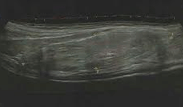
Patient is a 17-years-old obese male with a past medical history of well controlled Diabetes Mellitus who presented with progressive painless swelling in left thigh (Figure 1) since last six months. Patient reported that swelling was small initially, but it gradually increased in size. On physical examination, painless mass was noted that originated from Anterior Superior Iliac Spine (ASIS) and extended to lateral epicondyle of the left femur. Patient denied any weight loss, fever or other systemic symptoms. Lower extremity ultrasound was performed, and diagnosis of lipoma was confirmed (Figure 2). Lipoma excision was planned. Patient was placed under saddle anesthesia; long 12 cm vertical incision was made on the anterior aspect of left thigh and two well encapsulated masses were dissected in Figures 3(A and B). Specimen was sent for pathological evaluation and it confirmed two solitary lipomas which were yellowish, nodular with dull tan areas without any cellular atypia or abnormal infiltration and measured 16x11x8 cm and 16x15x8 cm respectively. Jackson Pratt (JP) drain was placed (Figure 4) which was removed after ten days. Wound care was suggested and sutures were removed after three weeks. The patient did not have any post operative deficits and was stable after removal of JP drain and suture removal. Post operatively patient was normal without any motor or sensory anomalies in the left lower extremity. Patient was also recommended to follow up with physical and occupational therapy for quick recovery. Keywords: Ascites, Inflammatory disease, Ovarian cancer, Pelvic tuberculosis | ||||||
| ||||||
| ||||||
| ||||||
| ||||||
|
DISCUSSION
| ||||||
|
Lipoma is benign soft tissue tumor that usually presents as slow growing, painless, well circumscribed mass varying in size in different parts of the body. Reported anatomical sites include joints, extra-dural with respect of spinal cord, breast, back and also other unusual locations like heart with or without septal involvement [1], [2], [3]. Numerous etiologies have been proposed for Lipoma. For instance, based on studies by Schoenmakers E. et al., aberrant genetic rearrangements on human chromosome 12 known as HMG1C have been known to cause solid benign lipomatous tumors [4]. In addition, hereditary conditions like Familial Multiple Lipomatosis have been known to present with multiple painless masses on trunk and extremities sparing the forehead with higher incidence in males as compared to females [5]. Other disorders include Dercum’s disease [6] and Madelung’s disease [7] with former being more commonly associated with obesity in post menopausal women and latter being associated with alcohol consumption. Lipomas can be characterized in several different ways [2]. These include solitary lipomas which are isolated, benign and well circumcised, angiomyolipomas which are tender subcutaneous masses which are somewhat firmer as opposed to the rubbery consistency and congenital diffuse lipomas which are composed of immature fat cells that occur on trunk and back. In addition, if lipomas are larger than ten cm, liposarcoma needs to be ruled out by ensuring lack of associated symptoms like weight loss [8]. In this case, we demonstrate a patient with an Intramuscular lipoma in the left lower extremity originating from ASIS, extending onto lateral epicondyle of the femur. It was interesting to see that despite such a huge size, patient did not have any sensory or vascular symptoms due to compression of neuro-vasculature located in that area. From a surgical stand point, during lipoma excision, care must be taken to ensure that muscle fibers and attachments are spared in order to prevent motor problems related to knee extension and hip flexion. Specifically injury to rectus femoris and vastus muscles is very common during cauterization which may cause muscular fibrosis that can present with motor symptoms post operatively. In addition, patient must be monitored for compartment syndrome secondary to tight facial closure. | ||||||
|
CONCLUSION
| ||||||
|
In summary, we have presented a case of a patient with a benign intramuscular lipoma that was successfully diagnosed and treated with surgical removal and excellent post-operative recovery. | ||||||
|
REFERENCES
| ||||||
|
||||||
SUGGESTED READING | ||||||
|
|
||||||


|
[HTML Abstract]
[PDF Full Text]
|
|
Author Contributions
Smit Shah – Substantial contributions to conception and design, Acquisition of data, Analysis and interpretation of data, Drafting the article, Revising it critically for important intellectual content, Final approval of the version to be published Praful Shah – Substantial contributions to conception and design, Acquisition of data, Analysis and interpretation of data, Drafting the article, Revising it critically for important intellectual content, Final approval of the version to be published |
|
Guarantor of Submission
The corresponding author is the guarantor of submission. |
|
Source of Support
None |
|
Consent Statement
Written informed consent was obtained from the patient for publication of this study. |
|
Conflict of Interest
Author declares no conflict of interest. |
|
Copyright
© 2018 Smit Shah et al. This article is distributed under the terms of Creative Commons Attribution License which permits unrestricted use, distribution and reproduction in any medium provided the original author(s) and original publisher are properly credited. Please see the copyright policy on the journal website for more information. |
|
|